How anterior imaging is revolutionising the eye care industry
14th February 2022
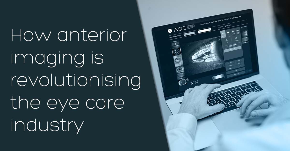
Photo documentation of the posterior segment of the eye is a long-established part of eye exams. It’s become routinely used by eye care professionals (ECPs) as a great way of educating patients about their eye health, as well as demonstrating the impact of their adherence to treatment plans. Only recently, however, have more ECPs begun to explore adopting anterior imagery into their offering as well.
The ability to show patients corneal edema, punctate staining or an epithelial defect is a great vehicle for educating patients, visually giving them a more substantial understanding of their condition as opposed to a verbal explanation. Anterior imaging also enables ECPs to track patients’ progress, with greater trust in these measurements thanks to the introduction of objective grading.
Examining the anterior segment is standard in the working day of any eye care professional and it has become standard practice for them to record findings using a recognised subjective grading system. However, these results are dependent on the ECPs interpretation – there is no guaranteed consistency between clinics and the professionals working within them. In fact, studies have shown reliability and accuracy between clinicians to be as low as 47%.
Thanks to revolutionary technology in the form of objective grading with AOS, grading accuracy and repeatability has shown to be as high as 99.8%. This is a significant boost in consistency that gives peace of mind for both the ECP and their patients.
An additional perk of incorporating anterior imaging into your clinic is the ability to lean on digital care delivery (tele-health) to offer safe and convenient care for patients. Nowadays, the vast majority of us have access to top-of-the-range smartphones/tablets equipped with highly sophisticated cameras. ECPs are already realising the benefits of utilising these devices for the patient and for the practice. A smartphone can be easily fitted with an adapter and attached to a slit lamp to create a digital slit lamp experience. In terms of offering the best delivery of eye care to enhance a practice, it’s also an alternative to parting with significant amounts of cash for a digital slit lamp. It’s therefore become apparent that a powerful imaging tool is in the hands of the majority of patients.
Combining anterior imaging with objective grading and digital care delivery, such as with AOS software, makes for an enhanced patient experience and improved time-efficiency for clinics. Patients are comfortable in the knowledge their data is safe and secure, all while getting reliable and educational treatment from the comfort of their own home. Eye care professionals, on the other hand, benefit from optimised chair time that maximises efficiency and revenue for the practice.