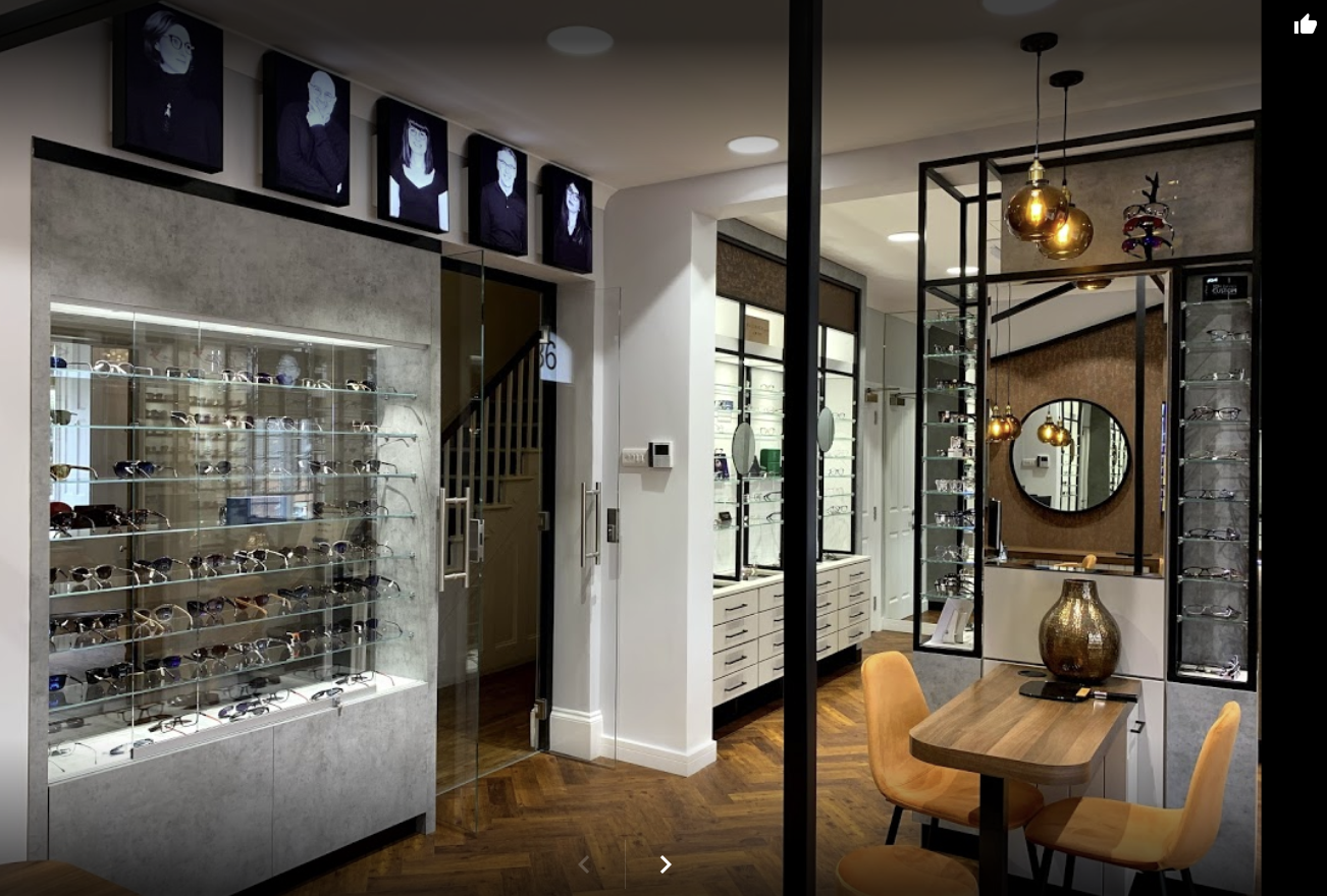
AOS Anterior in use during the Global Pandemic: Michelle Beach
17th June 2020

Michelle Beach of Park Vision Opticians recounts how her practice have experienced the pandemic and how AOS Anterior has helped them help their clients.
On March 23rd, we closed Park Vision’s doors to what we considered “normal” practice. From then on, our doors have been locked but for a buzz-in system. As a house practice, this was easy to do. We are away from the high street and have private parking right outside for our patients.
We have been busy keeping up to date with pandemic guidance and been in most days answering phone calls. We did the same as most other practices – updated our website and social media pages and tried to get all orders to patients as quickly as possible.
We had a lot of contact lens orders and I spent time driving glasses around Nottinghamshire -waving at patients from driveways and gates. Happily, our patients have been delighted with the efforts we made to get completed glasses and sunglasses out to them during the crisis.
However what about the emergencies, anxious patients with vision questions, or those with troublesome red eyes? Guidance from our governing bodies was to only see “face to face” if we absolutely had to, and to triage over the phone. Whilst that is great in theory, we know that one patient’s “agony” is another’s “a bit sore”!
What if we had missed something? What if what sounded like a mild case of hay fever was in fact something more serious? We are so used to being able to book our patients in and have the supporting armory of our diagnostic equipment to aid our clinical judgement. Without it we are a bit – excuse the pun – blind.
At Park Vision, I pride myself on having the best technology to support eyecare excellence. Our newly refurbished clinic incorporates new state-of-the-art teaching screens, video slitlamp and advanced dry eye technology. But this only works with a patient sitting in my clinic room!
I was aware of the remote imaging system from AOS from attending conferences earlier in the year. This tool suddenly seemed like no-brainer, offering peace of mind for me as a clinician and the quality of service my Park Vision patients expected.
A few emails and fantastic Zoom training from AOS Sales Director, Jeff Landucci, and we were live! This system has been essential in the new patient care we have had to adapt to over the last twelve weeks of lockdown. It is so simple – which patient doesn’t have access to a smart phone these days? That’s all that’s needed. A simple “selfie” using a mirror or a picture taken by a member of their household, and we can begin.
After a phone call, we send the patient an email giving clear steps on how to download an app and use it. The instructions are provided by AOS so I didn’t even have to construct a passage of text. Images sent through of the patient’s eye then arrive in the software inbox, and off you go. Using the software, you can crop and analyse as you see fit: highlight engorged vessels, areas of inflammation, bulbar redness – for any eye geek this is actually great fun!
If appropriate, you can then send the patient an annotated picture of their eye within a practice-branded email advising them to call for an appointment. Once again, this is all cleverly engineered into the software – you just fill in the blanks.
If a patient then needs an appointment, again the software is a triumph. Slit-lamp images can be brought to life, fluorescein staining can be enhanced, lesions demarcated….it will even count punctate staining for you.
To me, it is a valuable piece of technology to show a patient their eye issues. We all know a picture speaks a thousand words. It is a great educational tool and can produce a descriptive letter, including any enhanced images you have taken, for accurate referral. The images are stored enhancing record keeping.
But has the Anterior software actually worked? I have used this many times over the last few weeks. Patients are relieved that they are being “seen” without trying to describe their eye over the phone and are marvelling at the technology. Here are two specific cases I have dealt with during lockdown to illustrate how I have found using the Anterior software directly with my patients.
CASE 1: Corneal Ulcer
I had a call from a worried mother about her 18 year old daughter Grace – a long-standing patient. For the last few days Grace had complained of a sore eye. She was a high water content daily contact lens wearer. Grace suffered with hay fever and her mother assumed she was having a bad few days as the pollen count was high. That morning Grace was in a lot of pain, particularly when she woke and was struggling with the bright light.
I sent the AOS remote imaging instructions and quickly received the pictures of Grace’s eyes. It was obvious that her left eye was indeed very red, much more so than her right and that her symptoms warranted an appointment.

Grace arrived at Park Vision and slit-lamp images and staining showed a corneal ulcer. Grace and her mother were nervous about attending Eye Casualty but, following a phone call to my local hospital, and armed with the AOS images and letter, they headed off (see attached images).
Grace’s mum called me later that evening to say thank you. As she had the images included with the letter, the triaging nurse didn’t need to examine her. An ophthalmologist was called straight away and he treated very quickly, apparently marveling at the technology presented to him. Grace was “in and out” extremely quickly without the tedious waiting normally expected for this type of visit.

CASE 2: Dry Eye
I received another email from a very worried patient with sore, “stinging” eyes. Sohini was a visiting foreign university student. She emailed to say she wanted a face to face appointment as her eyes were very uncomfortable.
After explaining the guidelines and triage criteria I received Sohini’s eye pictures. This time, all looked fairly good. No particular redness or inflammation. The software analysis showed very few issues. On questioning, Sohini was worse when she was on her laptop and as her vision wasn’t affected, other than watery eyes. I came to the conclusion she was probably suffering with Dry Eyes. I made up a Dry Eye parcel for her, with clear instructions on a Dry Eye regime and she collected later that day from Park Vision.
An email three days later confirmed that her symptoms seemed to have alleviated.
Another three days later I received another email stating that she appeared to have gotten worse again. I spoke with her and, as she had been following the dry eye regime, I was concerned that I and my trusty AOS system had missed something. I booked her in for an appointment.

Sohini attended, grateful for an eye examination as she had been getting very worried. On presentation she still didn’t seem that bad. Slit-lamp images, staining and dry eye investigation revealed only mild symptoms and she was using the treatment. The issues had worsened when she increased her laptop time as she was trying to finish her thesis.
I took a quick look at her glasses which were 18 months old. She was wearing a myopic correction of R -0.25 and L -1.00. 6/6 in both eyes but also 6/6 unaided. With a +0.75 blur lens she was R 6/9 and L 6/7.5.
I explained to her that I thought her glasses correction was causing her issues. Unstable accommodation and binocular vision issues are known to be contributing factors among dry eye patients.
Sohini is booked for a more thorough eye examination this week. I actually think she will be hyperopic!
AOS Anterior did not let me down, it hadn’t missed anything and being able to show Sohini images of her eye on the screen reassured her enormously. It also helped her to realise that there might be other underlying causes.
Coming out of lockdown I will be continuing with the AOS imaging system. Whether in the clinic room or remotely, it has proven itself as a valuable asset in my consulting room.

Michelle Beach
