“Let me see!”: Allison Granger
12th August 2020
Last night after my son’s last baseball game, the family got together to take pictures with our little slugger. Immediately after the picture was taken, each member crowded around to see how it turned out, including my 17-month-old son. Yes, even my tiny tot grunted and pointed as if to say, “Let me see!”. We laughed about it. He already knows the drill. Take a look to make sure it’s a good one. It was, but it got me thinking about how image-obsessed our society is. Can you go one day without snapping a photo? I probably CAN, but I don’t. Smartphones are equipped with incredible cameras and I take advantage of that often. I snap pics to try to freeze time for a bit, or I use it to help me remember a product (like a good wine).
We see this trend in healthcare too, and for good reason. Images have tremendous value in documentation, monitoring, and patient education. We get copies of x-rays of broken bones to take home, vets can show us pictures of a dog’s teeth before and after cleaning, the gastroenterologist hands over a polaroid of the esophagus after an upper GI. There are countless examples, I’m sure you can think of your own.
Documentation is helpful for sure, but the patient education benefit of imaging is just as valuable and, in some cases, even more so. Patient education through images can lead to better understanding of the condition and better compliance to a treatment plan, which ultimately leads to better outcomes. Better yet, images can help validate the expense of the BEST treatment plan and not just the cheapest one.
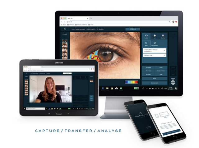
Think about this in terms of Ocular Surface and Dry Eye Disease assessment, treatment, and long-term patient management. Imagine a patient presents with anterior segment pathology. You take a picture, analyze the ocular surface and lids, and review what you see with the patient. With Anterior software, you can use an image taken from your slit lamp using the AOS companion app and enhance it to show the patient the graded objective output. That alone will get some ‘ooh’s and ‘ahh’s – or maybe even ‘eww’s and ‘arghhh’s!! Very few have ever seen their own eye in such detail. To really drive your point home, you also can tell them how it falls on the scale along with the percent vascularization. Now your patient is more educated and understanding of their eye condition, and likely thoroughly impressed by you. They also now have a number that they know they need to lower. They have seen with their own eyes what you see, and they are motivated to make it better.
You and the patient are on the same page, so it’s time to prescribe a treatment plan. Based on the diagnosis, there are two paths the patient can take: Option A is drops that are included in their health plan. Option B is a new in-office treatment that provides better outcomes but is an out of pocket expense. Armed with the information that the image and analysis provided, the patient can make a more informed choice, i.e. the best treatment plan.
When you have them back for a progress check, you take another image with analysis. You can track changes that the patient can see side by side (hopefully with more ‘ooh’s and less ‘eww’s) and either validate the plan or make any necessary changes. By educating the patient with images, you’ve improved outcomes, built trust, and I’ll go ahead and assume you’ve earned a patient for life!
If you haven’t thought about how anterior segment imaging plays in your practice, now’s the time to Let Them See.

Allison Granger
VP Of Business Development North America at AOS