Crossing Barriers with AOS: Dave Block
29th July 2020

Dave Block discusses crossing language barriers with imagery in contactology and optometry, including use of the AOS software.
Email, SMS, WhatsApp, messenger and many other apps developed in recent years have made communication easier than ever before. However, communication is one thing, but communicating correctly in a universal language that everyone can understand is another.
Grading scales are often used to communicate in contactology and optometry. A grading scale describes the condition and also the severity in degrees from 0 (normal) to 4 (severe).
The most famous grading scale in contactology is the Efron grading scale, shown below:
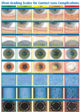
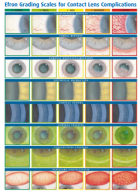
Until now, this was a grading scale that indicated the condition and severity by means of comparison images. It was therefore up to the specialist’s experience to correctly grade. This also immediately indicates the limitations and allows for human error. An experienced specialist will generally graduate differently from someone with less experience. This can result in incorrect communication between colleagues or ophthalmologists and subsequently adversely affect the treatment of the patient.
Objective grading with Artificial Intelligence
From the above it follows that correct grading, in which accuracy and the ability to grade in the same way, is essential. Here comes the advantage of software.
AOS Anterior
The AOS software allows images of the anterior eye segment to be analyzed in various ways. After that, the images can be easily interpreted and incorporated into the patient data system. This software shows even the most subtle changes between photos, allowing the progress of a condition to be accurately monitored. By working in this way it removes subjective error. An incorrect grading and interpretation based on individual assessment no longer plays a role. After all, it makes no difference now who takes the picture or sees the patient. The AOS software does the same for every user.
There are several eye disorders to be graded:
- Bulbar conjunctival redness
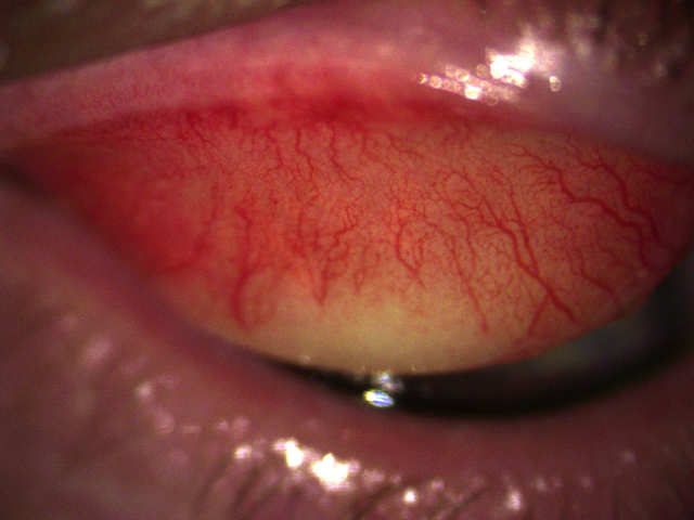
- Redness of the palpebral conjunctiva / eyelids
- Corneal staining
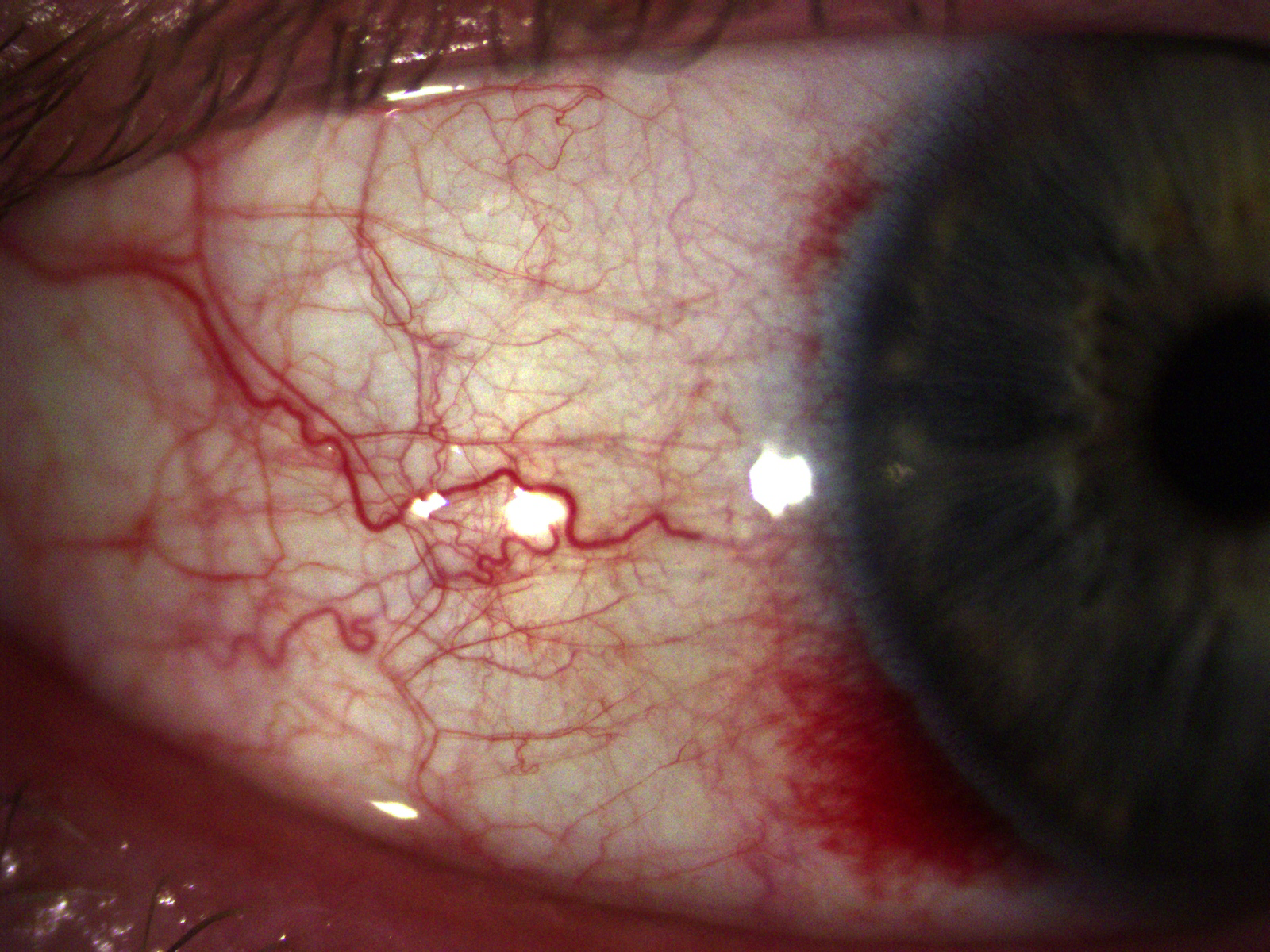
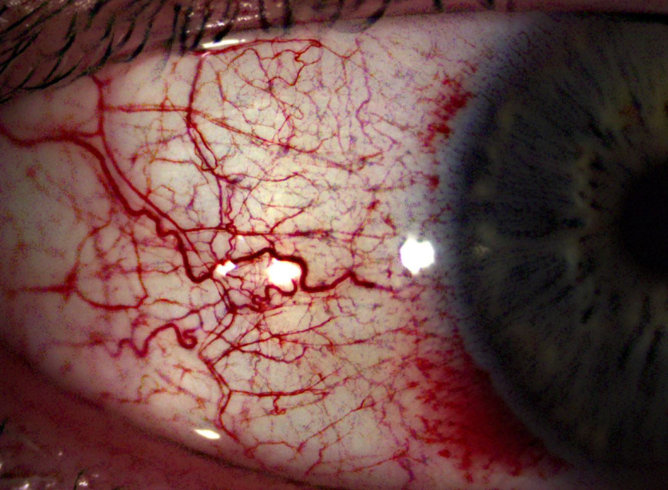
Above: Slit lamp photo and edited photo with clarification of the vessels
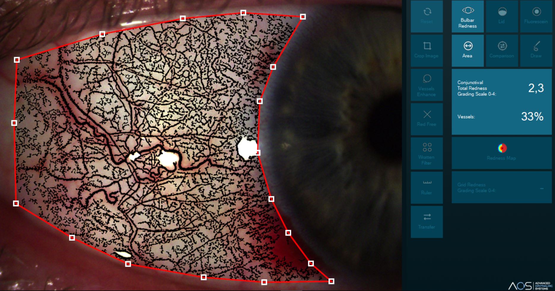
Above: Gradation conjunctival redness gr 2.3
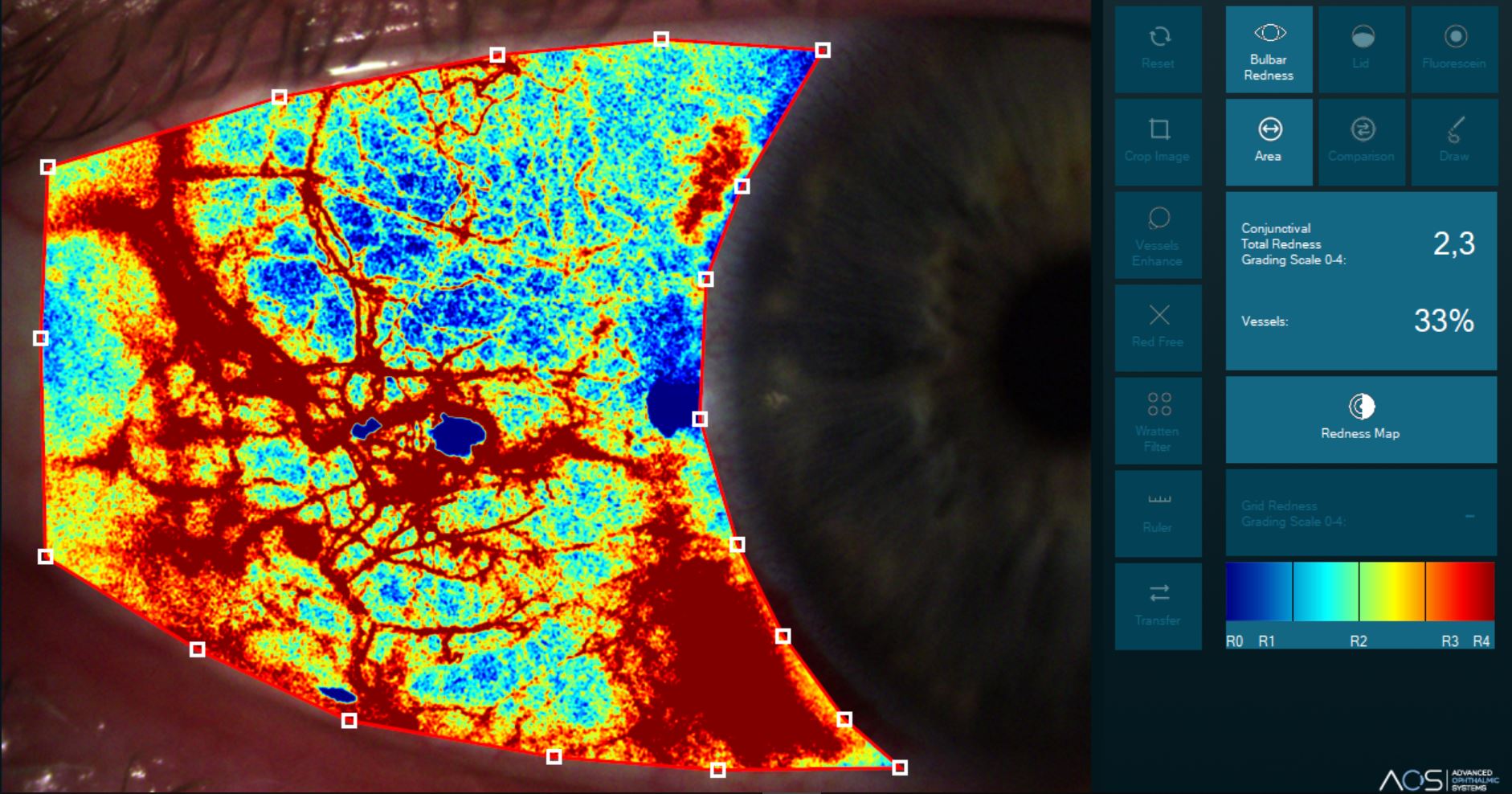
Above: “Heatmap” to clarify conjunctival redness.
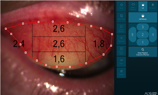
Above: Redness of the palpebral eyelid (= palpebral conjunctiva or eyelid) and associated grading.

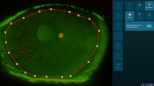
Above: Corneal staining images with and without gradation.
A nice extra is that a patient report can be made at the end, which can be included in a possible referral or on the status card.
After testing the AOS software, I believe it is easy to use and shows accurate and repeatable grades for key anterior eye segment conditions that are essential for the clinical management of patients. It indicates a bit of professionalism to the customer / patient and provides very accurate documentation for their own practice.

Dave Block studied at Gilde courses in Roermond. After completing his optics, he studied to become a contact lens specialist. His interest was always in solving contact lens related problems. This year he obtained his fellowship with the BCLA and obtained his specialization in dry eyes (OSCE). Dave works at Alcon as a Professional Affairs Manager and is involved in contact lens education in the form of a member of the construction committee as well as a member of the examination committee. In addition, he regularly teaches at the Department of General Practice (STOOHN) of the University of Maastricht and has his own company in ophthalmic diagnostics for people with diabetes.